Ultrasound: A Valuable Tool for Rheumatologists and Rheumatic Patients
June 10, 2024 | Rheumatic Disease

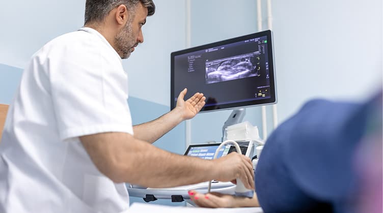
With the emergence of inexpensive portable ultrasound machines, ultrasound imaging has become a valuable tool for rheumatologists. In addition to the traditional physical exam, rheumatologists can now use ultrasound to see multiple targeted areas at once. Since ultrasound imaging can be performed in an office setting, this technology often cuts wait times and enables shared patient-provider decision making. Compared with most other imaging modalities, ultrasound increases patient safety, offers lower radiation levels, and is less expensive.
Musculoskeletal (MSK) ultrasound has been used in the field of rheumatology for more than two decades and has been used to detect joint effusions, synovitis, tendon pathologies, and bone erosions in a variety of diseases, including rheumatoid arthritis, psoriatic arthritis, gout and other inflammatory or crystalline arthropathies. Today, ultrasound is key to confirming a diagnosis and guiding treatment decisions. For instance, findings from ultrasound imaging may prompt a rheumatologist to increase or decrease corticosteroids requirement or even modify the immunosuppression base. Ultrasound imaging is also changing the way that rheumatologists administer treatments. For example, ultrasound-guided joint aspiration and injection have also been shown to be safer, less painful, and more accurate when compared to traditional approaches.
Ultrasound imaging has also been used in other fields of rheumatology, outside of musculoskeletal diseases. For example, vascular ultrasound with the examination of the temporal and axillary arteries can quickly assess patients suspected of having giant cell arteritis (GCA). Meanwhile, salivary gland ultrasound is used in Sjögren’s disease to aid in diagnosis, which often can be difficult. Muscle ultrasounds are currently being studied to help in faster diagnosis of inflammatory myopathies.
Simply put, ultrasound has changed the field of rheumatology and continues to evolve to provide a safer, quicker, and more accurate way for rheumatologists to care for rheumatic patients.

